World Radiography Day
Late on the evening of 8 November 1895, in a darkened laboratory at the University of Würzburg, the physicist Wilhelm Conrad Röntgen noticed a faint green glow on a screen coated with barium platinocyanide, several feet from a covered discharge tube that should not have been able to reach it. He had discovered a new kind of ray, one that passed straight through cardboard, wood and flesh but was stopped by bone and metal, and within weeks he had photographed the bones of his wife’s hand, her wedding ring hovering over the skeleton. World Radiography Day, held every year on 8 November, marks that evening — and honours the radiographers, the medical imaging professionals whose work turns Röntgen’s accident into diagnosis millions of times a day.
What World Radiography Day Marks
World Radiography Day is a professional and public-health observance dedicated to medical imaging and to the people who perform it: radiographers, also called radiologic technologists, along with the radiologists who interpret the images and the medical physicists who keep the equipment safe. The date is the anniversary of the discovery of the X-ray in 1895, and the day serves two purposes at once. It celebrates a technology that lets clinicians see inside the living body without cutting it open, and it raises the profile of a profession that patients rarely think about — the person behind the lead-lined door who positions the tube, sets the exposure and produces the image on which a diagnosis will rest. The observance is promoted globally by the International Society of Radiographers and Radiological Technologists together with the radiological societies of Europe and North America.
History
The discovery itself reads like a study in careful accident. Röntgen was experimenting with a Crookes tube, a sealed glass vessel through which an electric current produced a stream of what were then called cathode rays. He had wrapped the tube in black cardboard to block its visible light, and in the darkened room he saw a nearby fluorescent screen glowing anyway. Understanding at once that some unknown, penetrating radiation was responsible, he spent the following seven weeks working almost in secret, eating and sleeping in his laboratory. He called the new radiation “X” for unknown, and on 22 December he made the image that fixed the discovery in the public imagination: a fourteen-minute exposure of the left hand of his wife, Anna Bertha Ludwig, showing the bones and the dark shadow of her ring. She is said to have exclaimed that she had seen her own death.
Röntgen submitted his first paper, “On a New Kind of Rays”, on 28 December 1895, and the news moved with astonishing speed. Within weeks newspapers across Europe and America carried the story; within months physicians were using X-rays to locate bullets and set fractures. In 1901 Röntgen received the very first Nobel Prize in Physics, and in a gesture that shaped the field he refused to patent his discovery, insisting that it belonged to humanity, and donated his Nobel prize money to his university. The German word Röntgenstrahlen, “Röntgen rays”, is still the common term for X-rays across much of Europe.
The early enthusiasm carried a terrible cost that the day now quietly commemorates. Nobody understood that the rays were dangerous. Pioneers experimented on their own bodies, ran unshielded machines and stood in the beam for long periods, and many paid with burns, amputations and cancers. Clarence Dally, Thomas Edison’s assistant, worked with X-rays through the late 1890s, lost both arms to radiation injury and died in 1904, one of the first known martyrs of the field; Edison himself abandoned the research in horror. In Hamburg, a monument raised in 1936 in the garden of the St George hospital lists the names of radiologists and technicians from around the world who died from their work, and names have continued to be added. That memorial is the sober counterweight to the wonder of the discovery, and it is why the modern profession is built as much around radiation protection as around imaging.
Why It Matters
Medical imaging is now so deeply woven into medicine that its absence is almost unimaginable, and the humble X-ray remains the workhorse. A broken wrist, a suspected pneumonia, a swallowed coin, a dental abscess — all are read first on a radiograph, and the technology has branched into computed tomography, mammography, fluoroscopy and interventional procedures that thread catheters through the body under live imaging. The radiographer stands at the centre of all of it, balancing image quality against radiation dose according to the principle known as ALARA, “as low as reasonably achievable”, so that the diagnostic benefit always outweighs the small risk. World Radiography Day exists partly to make that invisible expertise visible, in the same spirit as the health observances that honour other unseen clinical professions such as the International Day of the Midwife.
The Profession Today
The person who takes an X-ray is a highly trained clinician, and one of the aims of the day is to correct the common assumption that they merely push a button. Radiographers hold degrees in medical imaging, understand anatomy and physics in equal measure, and make dozens of judgements for every image: how to position a frightened, injured patient; which exposure will show the pathology without over-irradiating; when an image is diagnostic and when it must be retaken. They staff emergency departments through the night, work in operating theatres alongside surgeons, and increasingly report on images themselves in some healthcare systems. The field has also become one of the frontiers of medical artificial intelligence, with algorithms now assisting in the reading of chest radiographs and mammograms, which has sharpened rather than dulled the debate about the irreplaceable role of the human in the loop.
The reach of the technology is deeply uneven across the world, and World Radiography Day increasingly draws attention to that gap. The World Health Organization has estimated that a large share of the global population still lacks access to any basic diagnostic imaging or radiography services at all, so that a fracture, a case of tuberculosis or an obstructed labour that would be imaged in minutes in a wealthy hospital goes unseen elsewhere. Campaigns tied to the day promote portable and solar-powered X-ray units, tele-radiography that lets images be read remotely, and the training of radiographers in low-resource countries, on the reasoning that a technology Röntgen gave freely to humanity ought in principle to reach all of it.
How It’s Marked
The day is observed mainly within hospitals, universities and professional bodies rather than in the streets. Radiography departments hold open days, lectures and skills demonstrations; imaging schools run competitions and awards for students; professional societies publish campaigns highlighting a chosen theme each year, from patient safety to the role of imaging in cancer care. Social media fills with historic images and with radiographers sharing the reality of a job that runs around the clock. Because the day sits so close to Röntgen’s own timeline, many events also fall within a wider Radiography Week, and it overlaps in purpose with the International Day of Radiology, which marks the same anniversary from the radiologists’ side of the profession.
Traditions and Symbols
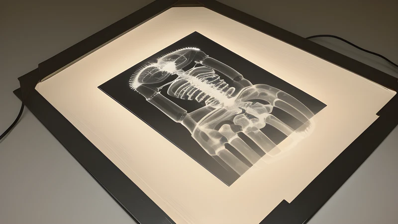
The enduring symbol of the day is Röntgen’s first medical radiograph, the ghostly hand with its ring, an image reproduced on posters and department walls wherever radiographers gather. The lead apron and the trefoil radiation-warning symbol stand for the safety culture that grew out of the early tragedies. And the skeleton hand itself has become a piece of visual shorthand for the whole idea of seeing through the surface of things, a motif that spread from the laboratory into art and popular culture within a year of the discovery.
Fun Facts
Röntgen never patented the X-ray and took no money from it, believing it should be freely available to all, which is one reason the technology spread so quickly. Within a year of the discovery, “X-ray photographs” had become a craze; shoe shops later installed fluoroscopes so customers could watch their own foot-bones wiggle inside a new shoe, a fad that ran into the 1950s before the radiation risk ended it. The element roentgenium, number 111 on the periodic table, is named in Röntgen’s honour, one of very few elements named for a scientist. And the very first clinical use of an X-ray in Britain came within months of the announcement, when doctors imaged a needle embedded in a woman’s hand — the diagnostic power of the ray was obvious to physicians almost from the first day they heard of it.
A Closing Reflection
There is a quiet lesson in the story that World Radiography Day preserves. A physicist noticed a glow he was not looking for, understood that he did not understand it, and spent seven weeks refusing to look away until he did. What began as an unexplained shimmer on a screen became, inside a single human lifetime, one of the foundations of modern medicine — and the price of learning to use it safely was paid in the health of the people who worked with it before anyone knew the danger. To mark the day is to hold both halves of that inheritance together: the wonder of seeing inside a living body, and the debt owed to the radiographers, past and present, who make that ordinary miracle safe.
Related Content